How NASH Progresses to Cirrhosis

The Cascade from Inflammation to Scar Tissue
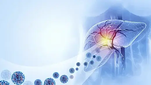
Nonalcoholic steatohepatitis (NASH), a severe form of nonalcoholic fatty liver disease (NAFLD), is a progressive condition that can lead to cirrhosis—a late-stage liver disease characterized by extensive fibrosis and impaired liver function. Understanding how NASH progresses to cirrhosis is crucial for early detection, intervention, and prevention of irreversible liver damage.
NASH results from chronic inflammation in the liver due to fat accumulation. Over time, the persistent injury triggers fibrogenesis, where the liver produces excess collagen, leading to scar tissue formation. Without timely intervention, this scarring can advance to cirrhosis, significantly increasing the risk of liver failure and hepatocellular carcinoma.
Unpacking the Stages of Liver Damage
1. Steatosis: The Initial Insult
Steatosis, or fatty liver, is the accumulation of triglycerides within hepatocytes. While often asymptomatic, it creates a metabolic environment that predisposes the liver to inflammation and oxidative stress. At this stage, lifestyle interventions such as weight loss and dietary modifications can reverse damage.
2. NASH: Transition from Fat to Inflammation
In NASH, the presence of fat leads to hepatocellular injury and immune activation. Inflammatory cytokines, including tumor necrosis factor-alpha (TNF-α) and interleukin-6 (IL-6), are released, perpetuating a cycle of damage. The liver’s regenerative response to this injury often results in early fibrosis.
3. Fibrosis: Laying the Groundwork for Cirrhosis
Fibrosis occurs when excess extracellular matrix (ECM) proteins, such as collagen, are deposited in the liver. This process begins as a protective response but becomes pathological as scar tissue accumulates. Fibrosis is classified into stages (F0 to F4), with F4 indicating cirrhosis.
For a detailed understanding of fibrosis staging, refer to the American Liver Foundation's guide to liver health.
The Mechanisms Driving NASH to Cirrhosis
Oxidative Stress
Increased fat accumulation elevates mitochondrial dysfunction and reactive oxygen species (ROS) production, which cause direct hepatocyte damage. The imbalance between ROS and the liver's antioxidant defenses accelerates tissue injury and inflammation.
Chronic Inflammation
Inflammatory cells such as macrophages and neutrophils infiltrate the liver, releasing pro-inflammatory cytokines. These molecules activate hepatic stellate cells (HSCs), the primary mediators of fibrogenesis.
Fibrogenesis
Activated HSCs produce collagen and other ECM components. Persistent activation results in excessive scar tissue that disrupts normal liver architecture, impeding blood flow and liver function.
Identifying Risk Factors for Disease Progression
Genetic Susceptibility
Variations in genes such as PNPLA3 and TM6SF2 are linked to a higher likelihood of developing NASH and progressing to cirrhosis. These genetic markers can help identify individuals at increased risk.
Metabolic Comorbidities
Conditions such as obesity, type 2 diabetes, and metabolic syndrome significantly increase the risk of NASH progression. Insulin resistance exacerbates lipotoxicity and inflammation, compounding liver injury.
Lifestyle Factors
Excessive caloric intake, sedentary behavior, and poor dietary habits contribute to disease progression. Conversely, adopting a Mediterranean diet and engaging in regular physical activity can mitigate risk.
Symptoms That Signal Cirrhosis
Cirrhosis often remains asymptomatic until significant liver function is lost. Early signs may include:
- Fatigue and weakness
- Unintentional weight loss
- Abdominal discomfort
- Jaundice (yellowing of the skin and eyes)
- Fluid retention leading to ascites
These symptoms should prompt immediate medical evaluation, as early intervention can slow or halt disease progression.
Diagnostic Approaches
Imaging
Techniques such as ultrasound, transient elastography (FibroScan), and magnetic resonance elastography (MRE) are non-invasive methods for assessing liver fat and fibrosis.
Blood Tests
Serum biomarkers like the NAFLD Fibrosis Score (NFS) and enhanced liver fibrosis (ELF) test provide insight into liver damage severity.
Liver Biopsy
A biopsy remains the gold standard for diagnosing NASH and assessing fibrosis stage. However, its invasive nature makes it a secondary option after non-invasive evaluations.
Treatment Strategies for NASH and Cirrhosis
Lifestyle Modifications
Weight loss is the cornerstone of treatment for NASH. Studies indicate that a 7-10% reduction in body weight can significantly improve liver histology, including resolution of NASH in some cases.
Pharmacological Interventions
Although no FDA-approved drugs specifically target NASH, several are under investigation, including obeticholic acid, elafibranor, and semaglutide. These medications aim to reduce inflammation, fibrosis, and liver fat.
Management of Cirrhosis
For patients with cirrhosis, the focus shifts to preventing complications such as variceal bleeding, hepatic encephalopathy, and ascites. Liver transplantation may be necessary in cases of decompensated cirrhosis.
Preventing Disease Progression
Regular Monitoring
Patients with NASH require regular follow-up to monitor disease progression. Non-invasive tools like transient elastography can track changes in fibrosis over time.
Addressing Comorbidities
Effective management of diabetes, hypertension, and hyperlipidemia is essential. Medications like statins and antidiabetic drugs may offer additional liver protection.
Understanding the Prognosis
While not all individuals with NASH progress to cirrhosis, those who do face a higher risk of liver-related mortality. Early diagnosis and intervention are critical to improving outcomes.
For more information on preventing liver disease progression, visit NIH Liver Disease Resources.
Conclusion
Understanding how NASH progresses to cirrhosis is essential for patients and healthcare providers alike. By recognizing risk factors, adopting preventative measures, and seeking early treatment, it is possible to halt or even reverse disease progression in its earlier stages. Timely intervention remains the cornerstone of managing this complex condition.
Share this article

Dr. Nico Pajes, MD
Dr. Nico Pajes is a board-certified internist and gastroenterologist with a focus on digestive health and internal medicine. See Full Bio.
-
1. Chalasani N, Younossi Z, Lavine JE, et al. "The Diagnosis and Management of Nonalcoholic Fatty Liver Disease: Practice Guidance." Hepatology, 2018.
-
2. Méndez-Sánchez N, et al. "Global Multi-Stakeholder Endorsement of the MAFLD Definition." Lancet Gastroenterol Hepatol, 2022.
-
3. Friedman SL. "Liver Fibrosis — From Bench to Bedside." J Hepatol, 2020.
-
4. Younossi Z, et al. "Nonalcoholic Steatohepatitis: Pathogenesis and Therapeutic Opportunities." Nat Rev Gastroenterol Hepatol, 2019.
-
5. Sanyal AJ, et al. "Mechanisms of Disease Progression in NAFLD and NASH." Gastroenterology, 2020.
-
6. Rinella ME. "Nonalcoholic Fatty Liver Disease: A Systematic Review." JAMA, 2015.
Genetic Mutations Linked to Fatty Liver Disease The interplay between genetics and disease has always been a subject of deep fascination.
Mastering Lifestyle for Fatty Liver Health The ping-pong match between one of my patients and his health had been ongoing for years. A software engineer...
The Role of Insulin Resistance in MAFLD Metabolic dysfunction-associated fatty liver disease (MAFLD), formerly known as non-alcoholic fatty liver...

You might enjoy more articles by
Dr. Nico Pajes, MD
 Disease
 Diets
Diets Recipes
 Supplements
 Management
 Calculators
 Quizzes
 Glossary


