The Role of Inflammation in NASH Development

A Key Driver in Non-Alcoholic Steatohepatitis
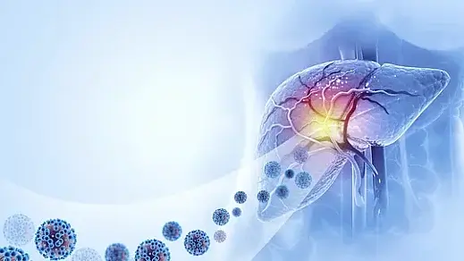
The role of inflammation in NASH development represents a critical area of hepatology research, highlighting the complex interplay between metabolic dysfunction and immune response in liver disease progression. Non-alcoholic steatohepatitis (NASH) emerges as a more severe form of non-alcoholic fatty liver disease (NAFLD), characterized by progressive inflammation that can lead to advanced fibrosis, cirrhosis, and hepatocellular carcinoma.
The Dual Nature of Inflammation in Liver Health
Inflammation is a natural immune response designed to protect the body from harm. However, chronic inflammation in the liver, as seen in NASH, becomes pathological. It involves the activation of hepatic immune cells, release of pro-inflammatory cytokines, and oxidative stress, all of which contribute to liver injury. These processes, when uncontrolled, lead to sustained liver damage and fibrosis.
What Triggers Inflammation in NASH?
Several factors contribute to the inflammatory response in NASH:
- Lipotoxicity: Excess lipid accumulation in hepatocytes triggers cellular stress and inflammatory pathways.
- Gut-Liver Axis Dysregulation: Increased intestinal permeability allows bacterial endotoxins to reach the liver, activating immune responses.
- Oxidative Stress: Reactive oxygen species (ROS) damage liver cells, promoting inflammation and fibrosis.
Emerging research highlights the significance of genetic predisposition and epigenetic modifications in amplifying these triggers.
Key Cellular Players in NASH Inflammation
Kupffer Cells
Kupffer cells, the liver’s resident macrophages, are pivotal in initiating and sustaining inflammation. When exposed to danger signals, such as lipotoxic molecules or bacterial endotoxins, they secrete pro-inflammatory cytokines like tumor necrosis factor-alpha (TNF-α) and interleukin-6 (IL-6).
Hepatocytes
Damaged hepatocytes release damage-associated molecular patterns (DAMPs), further activating immune cells and propagating inflammation.
Stellate Cells
Hepatic stellate cells play a dual role by promoting fibrosis in response to chronic inflammation. Activation of these cells leads to excessive extracellular matrix deposition, a hallmark of advanced NASH.
The Role of Cytokines and Chemokines
Inflammatory mediators like cytokines and chemokines orchestrate the immune response in NASH. Elevated levels of TNF-α, IL-1β, and IL-6 correlate with disease severity. These mediators not only recruit immune cells to the liver but also amplify oxidative stress and hepatocellular injury.
For an in-depth overview of cytokine pathways, read this comprehensive review by Nature.
Diagnostic Implications of Inflammation in NASH
Biomarkers
Emerging biomarkers reflecting inflammation and fibrosis offer non-invasive diagnostic tools for NASH. For instance:
- C-reactive Protein (CRP): Indicates systemic inflammation.
- Cytokeratin-18 Fragments: Reflect hepatocyte apoptosis and necrosis.
- Pro-inflammatory Cytokines: Elevated TNF-α and IL-6 levels suggest ongoing inflammation.
Imaging Techniques
Advanced imaging, such as magnetic resonance elastography (MRE), evaluates liver stiffness, indirectly reflecting inflammation and fibrosis. Combined with serological markers, it enhances diagnostic accuracy.
Therapeutic Approaches Targeting Inflammation
Lifestyle Interventions
Weight loss through dietary changes and increased physical activity remains the cornerstone of managing inflammation in NASH. A Mediterranean diet, rich in anti-inflammatory foods like omega-3 fatty acids, has shown significant benefits.
Pharmacological Strategies
Emerging drugs targeting inflammation in NASH include:
- TNF-α Inhibitors: Block pro-inflammatory pathways.
- FXR Agonists: Modulate bile acid pathways and reduce inflammation.
- Anti-oxidative Therapies: Combat oxidative stress to mitigate liver injury.
Future Directions and Therapeutic Implications
Recent advances in understanding the inflammatory mechanisms in NASH have opened new avenues for therapeutic intervention. Targeting specific components of the inflammatory cascade may provide more effective treatments while minimizing systemic effects. The development of combination therapies that address both metabolic and inflammatory aspects of the disease shows particular promise.
Conclusion
The role of inflammation in NASH development is multifaceted, involving a complex interplay of immune cells, cytokines, and molecular pathways. Addressing inflammation is pivotal for halting disease progression and improving patient outcomes. Ongoing research continues to refine our understanding and treatment of this challenging condition.
Share this article

Dr. Nico Pajes, MD
Dr. Nico Pajes is a board-certified internist and gastroenterologist with a focus on digestive health and internal medicine. See Full Bio.
-
1. Chalasani N, Younossi Z, Lavine JE, et al. The diagnosis and management of nonalcoholic fatty liver disease: Practice guidance from the AASLD. Hepatology, 2018.
-
2. Méndez-Sánchez N, Bugianesi E, Gish RG, et al. Global multi-stakeholder endorsement of the MAFLD definition. Lancet Gastroenterol Hepatol, 2022.
-
3. Rinella ME. Nonalcoholic fatty liver disease: A systematic review. JAMA, 2015.
-
4. Loomba R, Friedman SL. Mechanisms and disease consequences of nonalcoholic fatty liver disease. Cell Metabolism, 2020.
-
5. Eslam M, Sanyal AJ, George J. MAFLD: A consensus-driven proposed nomenclature for metabolic-associated fatty liver disease. Lancet Gastroenterol Hepatol, 2020.
-
6. Friedman SL, Neuschwander-Tetri BA, Rinella M, Sanyal AJ. Mechanisms of NAFLD development and therapeutic strategies. Nat Med, 2018.
How NASH Progresses to Cirrhosis Nonalcoholic steatohepatitis (NASH), a severe form of nonalcoholic fatty liver disease (NAFLD), is a progressive condition...
Weight Loss Quiz for Fatty Liver Weight loss is a cornerstone in managing nonalcoholic fatty liver disease (NAFLD), a condition affecting millions worldwide.
What Is a NAFLD Simulator? It was another bustling morning in the hospital when Dr. Rodriguez burst into the residents' lounge, coffee in hand, with...

You might enjoy more articles by
Dr. Nico Pajes, MD
 Disease
 Diets
Diets Recipes
 Supplements
 Management
 Calculators
 Quizzes
 Glossary


